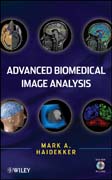
Medical diagnostics and intervention, and biomedical research rely progressively on imaging techniques, namely, the ability to capture, store, analyze, anddisplay images at the organ, tissue, cellular, and molecular level. These tasks are supported by increasingly powerful computer methods to process and analyze images. This text serves as an authoritative resource and self-study guideexplaining sophisticated techniques of quantitative image analysis, with a focus on biomedical applications. It offers both theory and practical examples for immediate application of the topics as well as for in-depth study INDICE: PREFACE. ACKNOWLEDGMENTS. CHAPTER 1: Image Analysis - A Perspective. 1.1. Introduction. 1.2. Main Biomedical Imaging Modalities. 1.3. BiomedicalImage Analysis. 1.4. Current Trends in Biomedical Imaging. 1.5. About this Book. 1.6. Chapter References. CHAPTER 2: Survey of Fundamental Image ProcessingOperators. 2.1. Statistical Image Description. 2.2. Brightness and Contrast Manipulation. 2.3. Image enhancement and restoration. 2.4. Intensity-based segmentation (thresholding). 2.5. Multidimensional thresholding. 2.6. Image calculations. 2.7. Binary image processing. 2.8. Biomedical Examples. 2.9. Chapter References. CHAPTER 3: Image Processing in the Frequency Domain. 3.1. The Fourier Transform. 3.2. Fourier-based filtering. 3.3. Other integral transforms: the discrete cosine transform and the Hartley transform. 3.4. Biomedical examples. 3.5. Chapter References. CHAPTER 4: Wavelet-Based Filtering. 4.1. Introduction to the wavelet transform and multiscale decomposition. 4.2. Wavelet-based filtering. 4.3. Comparison of frequency-domain analysis to wavelet analysis. 4.4. Biomedical examples. 4.5. Chapter References. CHAPTER 5: Adaptive Filtering. 5.1. Adaptive Filter Principles. 5.2. Adaptive Noise Reduction. 5.3. Adaptive filters in the frequency domain, adaptive Wiener filters. 5.4. Segmentationwith local adaptive thresholds and related methods. 5.5. Biomedical examples.5.6. Chapter References. CHAPTER 6: Deformable Models and Active Contours. 6.1. The Principle of Deformable Models. 6.2. Two-dimensional active contours (Snakes). 6.3. Three-dimensional active contours. 6.4. Live Wire techniques. 6.5. Biomedical Examples. 6.6. Chapter References. CHAPTER 7: The Hough Transform. 7.1. Detecting lines and edges with the Hough transform. 7.2. Detection of circles and ellipses with the Hough transform. 7.3. The Generalized Hough Transform. 7.4. The Randomized Hough Transform. 7.5. Biomedical Examples. 7.6. Chapter References. CHAPTER 8: Texture Analysis. 8.1. Statistical texture classification. 8.2. Texture classification with local neighborhood methods. 8.3. Frequency-Domain Methods for Texture classification. 8.4. Run-Lengths. 8.5. Other Classification Methods. 8.6. Biomedical Examples. 8.7. Chapter References. CHAPTER 9: Shape Analysis. 9.1. Cluster Labeling. 9.2. Spatial-Domain Shape Metrics. 9.3. Statistical Moment Invariants. 9.4. Chain Codes. 9.5. Fourier descriptors. 9.6. Topological Analysis. 9.7. Biomedical Examples. 9.8. Chapter References. CHAPTER 10: Fractal approaches to image analysis. 10.1. Self-similarity and the fractal dimension. 10.2. Estimation techniques for the fractal dimension in binary images. 10.3. Estimation techniques for the fractal dimension in grayscale images. 10.4. Fractal dimension in the frequency domain. 10.5. The local Hölder exponent. 10.6. Biomedical Examples. 10.7. Chapter References. CHAPTER 11: Image Registration. 11.1. Linear Spatial Transformations. 11.2. Nonlinear Transformations. 11.3. Registration Quality Metrics. 11.4. Interpolation Methods for Image Registration. 11.5. Biomedical Examples. 11.6. Chapter References. CHAPTER 12: Image storage, transport, and compression. 12.1. Image size, resolution, and depth. 12.2. Image Archiving, DICOM, and PACS. 12.3. Lossless Image Compression. 12.4. Lossy Image Compression. 12.5. Biomedical Examples.12.6. Chapter References. CHAPTER 13: Image Visualization. 13.1. Grayscale image visualization. 13.2. Color representation of grayscale images. 13.3. Contour Lines. 13.4. Surface rendering. 13.5. Volume visualization. 13.6. Interactive 3D rendering and Animation. 13.7. Biomedical Examples. 13.8. Chapter References. CHAPTER 14: Image Analysis and Visualization Software. 14.1. Image Processing Software - an Overview. 14.2. ImageJ. 14.3. Examples for image processing programs. 14.4. Crystal Image. 14.5. OpenDX. 14.6. Wavelet-related software.14.7. Algorithm Implementation. 14.8. Chapter References. APPENDIX A: Image Analysis with Crystal Image. APPENDIX B: Software provided on DVD.
- ISBN: 978-0-470-62458-6
- Editorial: John Wiley & Sons
- Encuadernacion: Cartoné
- Páginas: 550
- Fecha Publicación: 10/12/2010
- Nº Volúmenes: 1
- Idioma: Inglés
